
Emergency Neonatal Reconstruction, Neural Placode Preservation & Watertight Dural Closure
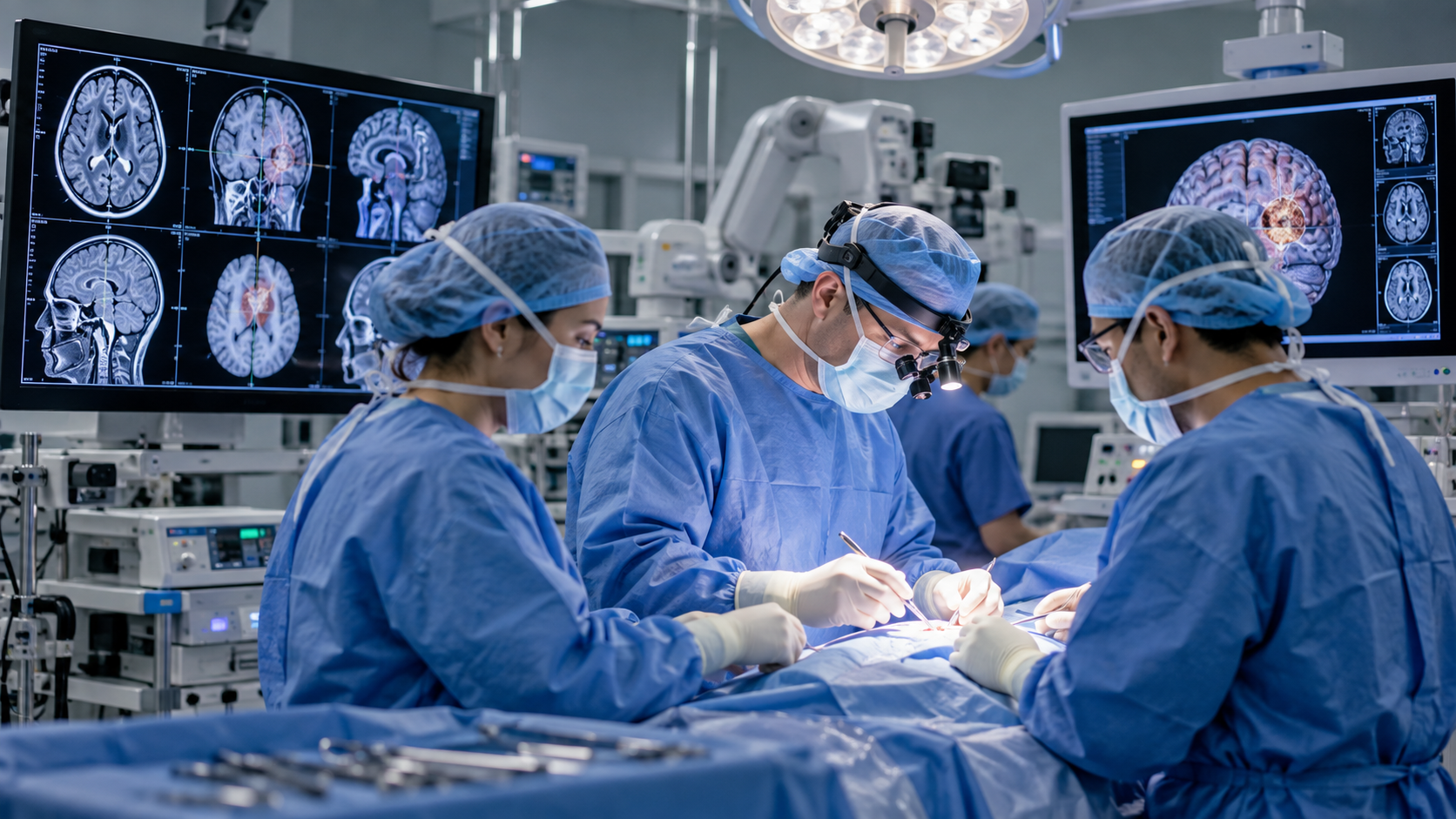
Meningomyelocele & Spina Bifida Neonatal Neurosurgery
Clinical Overview & Pathophysiology
Meningomyelocele (Myelomeningocele) is the most severe and clinically significant form of open Spina Bifida (dysraphism), resulting from failed primary neurulation and incomplete closure of the neural tube during embryonic days 26 to 28. In this condition, the posterior vertebral arches fail to fuse, and an open cystic sac containing exposed neural placode, malformed spinal cord tissue, and nerve roots protrudes through the dorsal defect into the external environment. This exposes delicate nervous tissue to chemical irritation from amniotic fluid and creates an immediate risk of fatal ascending bacterial meningitis following birth. In addition, over 85-90% of children with meningomyelocele have an associated Chiari II malformation and obstructive hydrocephalus. Dr. Ashok Kumar delivers urgent neonatal microsurgical repair within 24 to 48 hours of birth, focusing on neural placode preservation, multilayer watertight dural reconstruction, and proactive hydrocephalus management.
Clinical Classifications & Pathological Subtypes
- Open Myelomeningocele: The raw, exposed neural placode lies flush with the skin or atop a fluid-filled sac, actively leaking CSF into the environment.
- Meningocele: The cystic sac contains meninges and CSF only, with the functional spinal cord remaining inside the spinal canal (good neurological prognosis).
- Lipomyelomeningocele: A closed congenital dysraphism where a subcutaneous lipomatous mass infiltrates the spinal cord and tethers the conus medullaris.
- Rachischisis: The most extreme dysraphic defect where the neural tube completely fails to close over multiple spinal segments without an overlying membrane.
- Associated Chiari II Malformation: Downward herniation of cerebellar vermis, 4th ventricle, and medulla into the cervical canal causing stridor and swallowing apnea.
Key Symptoms & Clinical Presentation
!Emergency Red Flags & Immediate Surgical Indications
Active cerebrospinal fluid (CSF) leakage from the exposed placode, hypothermia, fever, irritability, or signs of ascending ventriculitis/meningitis. This demands immediate sterile saline dressing, intravenous antibiotics, and urgent neurosurgical repair within 24 to 48 hours.
Diagnostic & Neuro-Evaluation Workflow
Prenatal Ultrasound & Maternal Serum Alpha-Fetoprotein (MSAFP)
Elevated maternal AFP combined with mid-trimester anomaly scans identifying the "lemon sign" (frontal bone scalloping) and "banana sign" (cerebellar deformation).
Urgent Postnatal Brain & Whole-Spine 3T MRI
Detailed mapping of the neural placode anatomy, exclusion of split-cord malformations, confirmation of Chiari II malformation, and ventricular volume assessment.
High-Resolution Cranial & Renal Ultrasound
Bedside neonatal screening for ventriculomegaly, hydroureter, and renal parenchymal dysplasia.
Pre-Operative Neurological & Urodynamic Baseline
Assessment of voluntary lower extremity spontaneous movements and post-void residual urine volumes.
Advanced Treatments & Procedures by Dr. Ashok Kumar
Emergency Neonatal Microsurgical Placode Reconstruction
Under high operative magnification, the open neural placode is delicately dissected off the surrounding epithelialized zona epithelioserosa and tubularized.
Multilayer Watertight Dural Closure
Reconstitution of the thecal sac using native dura mater (or synthetic collagen matrix graft) to achieve a hermetic, watertight barrier against CSF leaks.
Tension-Free Fascial & Cutaneous Flap Closure
Mobilization of extensive lumbodorsal myofascial flaps and relaxing skin incisions (Z-plasty or bipedicled flaps) to ensure durable, full-thickness coverage.
Concurrent or Staged VP Shunt / ETV
Placement of an infant programmable VP shunt or Endoscopic Third Ventriculostomy to manage concomitant progressive hydrocephalus.
Pediatric Tethered Cord Release
Elective secondary micro-dissection of filum terminale adhesions if neurological or urological deterioration emerges during childhood growth spurts.
Post-Operative Recovery & Long-Term Prognosis
The neonate is cared for in the Neonatal Intensive Care Unit (NICU) in a prone or lateral position for 5 to 7 days to eliminate pressure on the repair site. Dr. Ashok Kumar coordinates multidisciplinary lifelong care involving pediatric urology (clean intermittent catheterization), pediatric orthopedics, and developmental physiotherapy.
Frequently Asked Questions (Clinical FAQs)
Authoritative, medically verified answers to critical clinical questions regarding symptoms, surgical safety, recovery timelines, and long-term prognosis.
Why must a meningomyelocele be surgically closed within the first 24 to 48 hours of life?
Will surgery restore movement in a baby who is born with paralyzed legs?
How are hydrocephalus and Chiari II malformation managed in babies with spina bifida?
What is Tethered Cord Syndrome and will a child who had meningomyelocele surgery need another operation later?
Can a woman reduce the risk of having a baby with spina bifida in future pregnancies?
Spine Surgery & Spinal Disorders
Why Choose Dr. Ashok Kumar
Emergency 24 Hours Dr. Ashok Kumar
Neuro-Spine OPD Location
Direct Consultation & Emergency
Email: dr.ashokkumar4108@gmail.com
Consultation Hours
Emergency Neuro-Trauma: 24/7 Open