Microdiscectomy, Endoscopic Spine Decompression & Advanced Cervical/Lumbar Solutions
Disc Problems, Sciatica & Minimally Invasive Spine Surgery
Clinical Overview & Pathophysiology
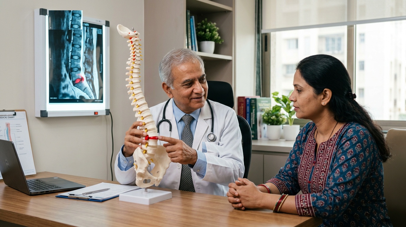
Intervertebral disc pathology is one of the most common causes of debilitating pain and neurological deficit, affecting the cervical (neck) and lumbar (lower back) spine. Degenerative disc disease begins with dehydration of the nucleus pulposus and tears in the surrounding annulus fibrosus, culminating in disc bulging, herniation (slipped disc), extrusion, or sequestration. When displaced disc material compresses adjacent exiting spinal nerve roots or the central thecal sac, it produces severe radiating pain (sciatica or cervical radiculopathy), sensorimotor weakness, and neurogenic claudication. Dr. Ashok Kumar specializes in advanced Minimally Invasive Spine Surgery (MISS), including tubular microdiscectomy, full-endoscopic discectomy, and anterior cervical discectomy with fusion (ACDF), restoring mobility through sub-centimeter incisions.
Clinical Classifications & Pathological Subtypes
- Lumbar Disc Herniation (L4-L5, L5-S1): Posterolateral disc rupture compressing the descending L5 or S1 nerve roots, generating severe sciatica down the posterior leg to the foot.
- Cervical Disc Herniation (C5-C6, C6-C7): Cervical root impingement producing agonizing shoulder, arm, and finger pain, numbness, and biceps/triceps weakness.
- Lumbar Spinal Canal Stenosis: Narrowing of the central spinal canal by hypertrophied facet joints, thickened ligamentum flavum, and diffuse disc bulges causing neurogenic claudication.
- Cervical Spondylotic Myelopathy (CSM): Progressive spinal cord compression in the neck resulting in clumsiness of hands, unsteady spastic gait, and balance loss.
- Lumbar Spondylolisthesis: Anterior slip of one vertebra over another due to pars interarticularis defects (isthmic) or facet joint degeneration, creating dynamic nerve impingement.
Key Symptoms & Clinical Presentation
!Emergency Red Flags & Immediate Surgical Indications
Cauda Equina Syndrome: Sudden onset of bilateral leg sciatica, numbness in the groin and buttocks (saddle anesthesia), difficulty passing urine (retention), or loss of bowel control. Cauda Equina Syndrome is an absolute surgical emergency requiring decompression within 24 hours to prevent permanent incontinence and sexual dysfunction.
Diagnostic & Neuro-Evaluation Workflow
High-Resolution Lumbar / Cervical 3T MRI
Sagittal and axial T2-weighted sequences demonstrating the precise anatomical grade of disc extrusion, canal diameter, and thecal compression.
Dynamic Flexion-Extension Spine Radiography
Weight-bearing dynamic stress X-rays assessing hypermobility, segmental instability, or dynamic spondylolisthesis.
Electromyography (EMG) & Nerve Conduction Velocity (NCV)
Electrophysiological testing confirming acute or chronic radiculopathy and ruling out peripheral neuropathies (e.g., diabetic neuropathy or peroneal nerve palsy).
High-Resolution Non-Contrast Spine CT
Detailed delineation of hard osteophytic bone spurs, ossification of the posterior longitudinal ligament (OPLL), and vacuum disc phenomenon.
Advanced Treatments & Procedures by Dr. Ashok Kumar
Tubular Minimally Invasive Microdiscectomy (MISS)
Through a 1.5 cm skin incision and tubular muscle-splitting dilators, an operative microscope is utilized to extract the herniated disc fragment while sparing back muscles.
Full-Endoscopic Lumbar Discectomy
Ultra-minimally invasive transforaminal or interlaminar endoscopic spine decompression performed through an 8-mm port under continuous fluid irrigation.
Anterior Cervical Discectomy & Fusion (ACDF)
Removal of herniated cervical disc through a natural anterior neck skin crease, followed by insertion of an anatomical PEEK cage packed with osteoinductive graft.
Cervical Artificial Disc Replacement (Arthroplasty)
Motion-preserving implant replacing damaged disc, maintaining natural neck kinematics and protecting adjacent spinal segments from accelerated degeneration.
Minimally Invasive Transforaminal Lumbar Interbody Fusion (MIS-TLIF)
Percutaneous screw fixation and interbody cage placement for lumbar instability and spondylolisthesis with minimal tissue disruption.
Post-Operative Recovery & Long-Term Prognosis
Following minimally invasive microdiscectomy, patients are encouraged to walk within 4 to 6 hours of surgery. Over 90% of patients are discharged either the same evening or the following morning. Return to non-strenuous desk work occurs within 10 to 14 days, supported by targeted core-stabilization physiotherapy.
Frequently Asked Questions (Clinical FAQs)
Authoritative, medically verified answers to critical clinical questions regarding symptoms, surgical safety, recovery timelines, and long-term prognosis.
When is surgery necessary for a slipped disc instead of continuing medication and rest?
What are the main advantages of Minimally Invasive Microdiscectomy over traditional open spine surgery?
What is Cauda Equina Syndrome and why is it considered a neurosurgical emergency?
What is the risk of a slipped disc recurring after surgery?
Can artificial disc replacement be performed instead of spinal fusion in the neck?
Spine Surgery & Spinal Disorders
Why Choose Dr. Ashok Kumar
Emergency 24 Hours Dr. Ashok Kumar
Neuro-Spine OPD Location
Direct Consultation & Emergency
Email: dr.ashokkumar4108@gmail.com
Consultation Hours
Emergency Neuro-Trauma: 24/7 Open