Foramen Magnum Decompression, C1-C2 Goel-Harms Realignment & Tethered Cord Release
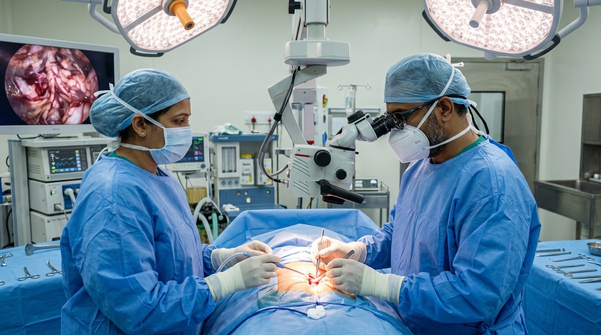
Congenital Spine Anomalies, Chiari Malformation & CV Junction Surgery
Clinical Overview & Pathophysiology
Congenital spine defects comprise structural anomalies arising from embryological errors in segmentation, formation, or blastogenesis of the axial skeleton and neural tube. Prominent among these are Craniovertebral (CV) Junction Anomalies—including Basilar Invagination, Atlantoaxial Dislocation (AAD), and Os Odontoideum—which cause direct compression of the cervicomedullary junction and upper cervical cord. Closely linked is Chiari Malformation Type I (downward herniation of cerebellar tonsils > 5 mm below the foramen magnum) frequently associated with Syringomyelia (fluid-filled syrinx cavity inside the spinal cord). Other congenital dysraphisms include Tethered Cord Syndrome and Congenital Scoliosis. Dr. Ashok Kumar delivers world-class micro-neurosurgical decompression, CV junction realignment, Goel-Harms C1-C2 screw fixation, and detethering procedures.
Clinical Classifications & Pathological Subtypes
- Chiari Malformation Type I & Syringomyelia: Cerebellar tonsillar ectopia through the foramen magnum impeding normal CSF pulsation, resulting in a progressive intramedullary fluid cavity (syrinx).
- Atlantoaxial Dislocation (AAD) / Subluxation: Instability between the atlas (C1) and axis (C2) vertebrae compressing the brainstem, classified into reducible and irreducible forms.
- Basilar Invagination & Platybasia: Upward protrusion of the odontoid process into the foramen magnum, compromising the lower brainstem and lower cranial nerves.
- Tethered Cord Syndrome & Split Cord Malformations (Diastematomyelia): Abnormal anchoring of the conus medullaris by a short, thickened filum terminale or osseous/fibrous spur bisecting the spinal cord.
- Congenital Scoliosis / Hemivertebrae: Structural spinal curvature resulting from failure of vertebral formation (hemivertebra) or failure of segmentation (unsegmented bar).
Key Symptoms & Clinical Presentation
!Emergency Red Flags & Immediate Surgical Indications
Acute respiratory arrest, sudden loss of gag reflex with aspiration pneumonia, rapid quadriplegia following minor neck trauma, and downbeat nystagmus. These indicate critical cervicomedullary junction strangulation demanding emergency surgical intervention.
Diagnostic & Neuro-Evaluation Workflow
3T Cranio-Cervical MRI with Phase-Contrast CSF Flow Cine
Quantifies tonsillar ectopia below the McRae line, delineates the extent of syringomyelia, and visualizes CSF flow jet turbulence across the foramen magnum.
Dynamic Multi-Slice CT of CV Junction with 3D Bone Rendering
Precision craniometry measuring Chamberlain's, McGregor's, and Wackenheim's lines, clival-canal angle, and facet joint orientation.
Whole-Spine MRI Screening
Mandatory imaging of the entire neuraxis to detect occult syrinx cavities, diastematomyelia bony spurs, and low-lying conus medullaris.
Flexion-Extension Cervical Radiographs
Assessing reducible vs fixed atlantoaxial dislocation (AAD) under physician supervision.
Advanced Treatments & Procedures by Dr. Ashok Kumar
Foramen Magnum Decompression (FMD / PFD)
Suboccipital craniectomy, C1 posterior arch resection, arachnoid-preserving micro-dissection, and expansile duroplasty restoring normal cisterna magna CSF dynamics.
C1 Lateral Mass - C2 Pedicle Screw Fixation (Goel-Harms Technique)
The gold-standard motion-preserving rigid fixation system providing immediate reduction, joint distraction, and arthrodesis for Atlantoaxial Dislocation.
Endoscopic Endonasal / Transoral Odontoidectomy
Resection of irreducible retroflexed odontoid bone causing ventral brainstem compression, followed by posterior occipitocervical fusion.
Microsurgical Release of Tethered Cord
Laminotomy, division of the thickened, fibrotic filum terminale, and excision of bony/fibrous diastematomyelia spurs.
Hemi-epiphysiodesis & Hemivertebra Excision
Surgical resection of malformed wedge-shaped vertebrae to halt progressive congenital scoliosis.
Post-Operative Recovery & Long-Term Prognosis
Following foramen magnum decompression, exertional occipital headaches generally resolve within days, and follow-up MRI scans at 6 to 12 months document dramatic shrinkage of the spinal syrinx. Patients undergoing C1-C2 screw stabilization wear a supportive cervical collar for 6 to 8 weeks until solid bony union is radiographically confirmed.
Frequently Asked Questions (Clinical FAQs)
Authoritative, medically verified answers to critical clinical questions regarding symptoms, surgical safety, recovery timelines, and long-term prognosis.
What is a Chiari Malformation and how does it create a fluid cavity (syrinx) inside the spinal cord?
What are the symptoms of Basilar Invagination and Atlantoaxial Dislocation (AAD)?
How does the Goel-Harms C1-C2 screw fixation technique stabilize the neck safely?
What are the classic signs of Tethered Cord Syndrome in children and teenagers?
Can syringomyelia heal completely after Foramen Magnum Decompression surgery?
Spine Surgery & Spinal Disorders
Why Choose Dr. Ashok Kumar
Emergency 24 Hours Dr. Ashok Kumar
Neuro-Spine OPD Location
Direct Consultation & Emergency
Email: dr.ashokkumar4108@gmail.com
Consultation Hours
Emergency Neuro-Trauma: 24/7 Open